Why Skin Cancer Screening Matters
Skin cancer is the most commonly diagnosed cancer in the United States, according to the Centers for Disease Control and Prevention (CDC). The encouraging news is that when detected early — particularly for melanoma, the most dangerous type — survival rates are significantly higher. The challenge is that many people only see a dermatologist after a change has become impossible to ignore.
Preventive skin exams shift that dynamic. Rather than waiting for symptoms, scheduled professional checks create an opportunity to identify abnormal growths before they progress. Understanding when those visits are genuinely necessary, versus when watchful self-monitoring is sufficient, helps you make informed decisions about your own care.
Recognizing the Warning Signs: The ABCDE Rule
Dermatologists use the ABCDE framework to help both clinicians and patients evaluate moles and lesions. Each letter corresponds to a characteristic that may signal a problem:
- Asymmetry: One half of the mole doesn't match the other.
- Border: Edges are irregular, ragged, notched, or blurred.
- Color: The lesion contains multiple shades — brown, black, red, white, or blue.
- Diameter: The spot is larger than 6 millimeters (roughly the size of a pencil eraser), though melanomas can be smaller.
- Evolving: Any change in size, shape, color, or new symptoms such as bleeding or itching.
The "E" for Evolving is often considered the most critical. A mole that has been stable for years but suddenly changes warrants prompt professional evaluation, regardless of whether it meets the other criteria.
Age-Based and Risk-Based Screening Guidance
There is no single universal screening schedule for skin cancer endorsed by all major health organizations, but clinical practice offers practical benchmarks. For average-risk adults, most dermatologists suggest beginning annual full-body skin exams around age 40, or earlier if risk factors are present.
Higher-risk individuals — including those with fair skin, light eyes, a history of blistering sunburns, substantial cumulative sun exposure, a personal or family history of skin cancer, or a large number of moles — may benefit from starting screenings in their 20s or 30s and repeating them more frequently. People who have had any prior skin cancer diagnosis are typically monitored every three to six months, depending on the type and stage.
It's worth noting that the U.S. Preventive Services Task Force (USPSTF) has stated that current evidence is insufficient to recommend routine screening for the general asymptomatic population. However, this does not mean screenings lack value — it reflects limitations in the research, not a conclusion that exams are harmful. Talking with your primary care provider or dermatologist about your personal risk profile is the most reliable way to determine an appropriate schedule.
How to Conduct a Monthly Self-Exam
Self-exams complement professional care by helping you detect new or changing lesions between appointments. A thorough self-exam takes about 10 minutes and should be done monthly in good lighting using a full-length mirror and a hand mirror for hard-to-see areas.
Examine the entire body systematically: face, scalp (use a comb or hair dryer to part hair), neck, chest, arms, hands, torso, back, legs, feet — including the soles and between the toes. Don't overlook areas less exposed to the sun; melanoma can develop anywhere, including beneath fingernails and on the scalp.
Keep a simple log or photograph suspicious spots so you can track any evolution over time. If you notice anything that fits the ABCDE criteria or simply feels "different," schedule a professional evaluation rather than waiting for your next routine appointment.
What to Expect During a Full-Body Skin Exam
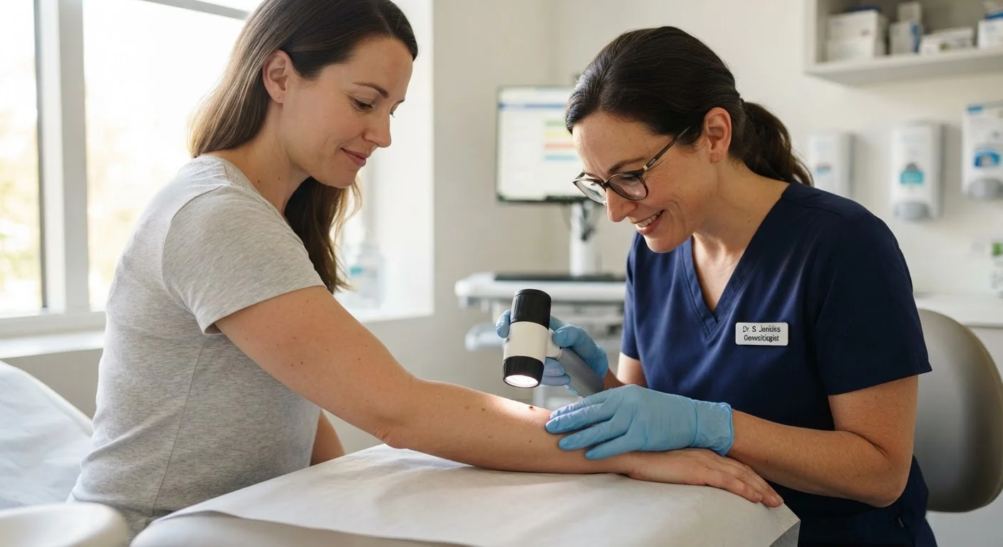
A full-body skin exam with a board-certified dermatologist typically takes 15 to 30 minutes. You'll be asked to undress to allow examination of all skin surfaces, including the scalp, genitals, and between the toes — areas patients often overlook themselves. The dermatologist may use a dermoscope, a handheld device that magnifies skin structures and reveals patterns invisible to the naked eye.
If a suspicious lesion is found, the dermatologist may photograph it for monitoring, perform a biopsy (removing a small tissue sample for lab analysis), or refer you to another specialist. A biopsy is the only definitive way to diagnose skin cancer. The procedure is typically quick and performed under local anesthesia in the office.
Bring a list of any spots you've noticed or are concerned about, and don't hesitate to point them out even if the dermatologist has already passed over that area. Your observations matter.
This article is for general informational purposes only and does not constitute medical advice. Always consult a qualified healthcare professional for personal medical decisions, new or changing skin lesions, or any other health concerns.